Skin Cancer Application for SmartPhone
The problem:
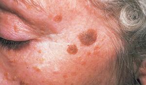
Some of the more recent information about spotting skin cancer has pointed out that one of the most important things to know when searching skin for cancer is how things have changed with time. If spots or growths have changed, they deserve attention. Many of us as we get older have many different spots on our skin -- all over our body. It is very difficult for a dermatologist to spot a problem, even if all of the spots are looked at carefully. For individuals like myself, who have seborrheic keratosis, searching all of our skin can take a long time. There are too many objects or discolorations for any doctor to be able to remember, and too many for most patients to recognize changes, since changes occur slowly over time.
The Solution:
Since so many people now have smart phones, I believe that an application could be built that would allow us (patients) to scan our own skin, map it, record it and then do automatic comparisons from time to time to look and identify changes. The patient would perform a self scan of their body with the camera on their smartphone, where the resulting files would be encrypted and sent to a secure processor that would stitch the photos together, form comparisons and flag changes. The resulting images and flags would be then made available to the patients dermatologist. When the patient came in for an appointment, the doctor would be cued for possible areas of concern.
Advantages:
-
Patient does own scanning in privacy of own home
-
Could cut down on time spent by dermatologist and improve accuracy
-
Will help flag those subtle changes that could indicate skin cancer problems
-
Forms a permanent record of a patient’s skin and shows changes with time
-
When some objects are removed from patients by surgery or freezing, it allows doctors to clearly see if they come back
-
This permanent record database could provide information for researchers trying to study types of skin cancers, discolorations, and growths
Patient does own scanning in privacy of own home
Could cut down on time spent by dermatologist and improve accuracy
Will help flag those subtle changes that could indicate skin cancer problems
Forms a permanent record of a patient’s skin and shows changes with time
When some objects are removed from patients by surgery or freezing, it allows doctors to clearly see if they come back
This permanent record database could provide information for researchers trying to study types of skin cancers, discolorations, and growths
How it would work for patient:
Patient would download the application, based upon doctor's instruction, and then find a place with good lighting and follow vocal instructions provided by the application. Instructions would be like this:
-
While holding the phone in your right hand, Press the plus ( +) button on your phone and slowly pass the camera over your left arm starting at the shoulder down to the tips of your fingers. When done press the minus (-) sign button Your scan should take about 5 seconds from start to finish.
-
Now do the same thing for the underside of your arm from your left armpit down to the tips of your fingers. Press plus sign to start and minus sign to stop
-
Finally, turn your left hand so palm is up, and scan under the arm past the elbow down to your wrist.
The whole process could take as long as a half hour to complete scanning the whole body.
While holding the phone in your right hand, Press the plus ( +) button on your phone and slowly pass the camera over your left arm starting at the shoulder down to the tips of your fingers. When done press the minus (-) sign button Your scan should take about 5 seconds from start to finish.
Now do the same thing for the underside of your arm from your left armpit down to the tips of your fingers. Press plus sign to start and minus sign to stop
Finally, turn your left hand so palm is up, and scan under the arm past the elbow down to your wrist.
What the application would do:
-
Create a file for each of the scans along with metadata for the scan
-
Verify that the lighting for the imagery is adequate for recording
-
Make sure that the scan wasn’t too fast or too slow.
-
Encrypt the file for privacy based upon keys provided by the server
-
Uplink the file to a server
Create a file for each of the scans along with metadata for the scan
Verify that the lighting for the imagery is adequate for recording
Make sure that the scan wasn’t too fast or too slow.
Encrypt the file for privacy based upon keys provided by the server
Uplink the file to a server
What the server and software will do:
-
Capture all of the files uploaded and do a quality check to make sure the data is processable and is complete.
-
Report in real time any errors, to allow the patient to repeat a scan if required
-
Real time errors could include corrupted files, missing files etc
-
Capture the patients metadata from the uploaded file and determine if there is a doctor to bill.
-
Stitch the files together to provide continuous skin coverage and align it based upon skin patterns (similar to stitching together landscape photos)
-
Determine if the patient has a file from a previous scan. If so, perform a comparison of the two files and identify changes based upon criteria developed from clinical tests:
-
Tolerance for how much change is enough to be flagged, to prevent false alarms
-
Tolerance could be based upon shape, size, and color of marking or object on skin
-
If too many differences, software could self-adjust some tolerances and report the situation. Could also determine if problem could be due to a photo registration/stitching problem
-
Doctor may want to set a specific tolerance for change, depending upon the patient's age and skin type
-
Extra filters may need to be enabled for patients who have other forms of skin problems, such as keratosis pilaris , hydrocarbon keratosis, or eczema.
-
Inform the doctor’s office that a patient’s file has been processed and is available for review.
Capture all of the files uploaded and do a quality check to make sure the data is processable and is complete.
Report in real time any errors, to allow the patient to repeat a scan if required
Real time errors could include corrupted files, missing files etc
Capture the patients metadata from the uploaded file and determine if there is a doctor to bill.
Stitch the files together to provide continuous skin coverage and align it based upon skin patterns (similar to stitching together landscape photos)
Determine if the patient has a file from a previous scan. If so, perform a comparison of the two files and identify changes based upon criteria developed from clinical tests:
- Tolerance for how much change is enough to be flagged, to prevent false alarms
- Tolerance could be based upon shape, size, and color of marking or object on skin
- If too many differences, software could self-adjust some tolerances and report the situation. Could also determine if problem could be due to a photo registration/stitching problem
- Doctor may want to set a specific tolerance for change, depending upon the patient's age and skin type
- Extra filters may need to be enabled for patients who have other forms of skin problems, such as keratosis pilaris , hydrocarbon keratosis, or eczema.
Inform the doctor’s office that a patient’s file has been processed and is available for review.
What the doctor’s interface will do:
-
Doctor’s credentials and billing information will be validated
-
Doctor will have a screen display showing the flagged locations where significant changes have been identified
-
When doctor clicks on that spot on the body, the display will show a split-screen display with previous scan and current scan. Doctor could opt for an overlay display and could click to see before and current scan.
-
Doctor could also look back in archives if patient had multiple scans on file and perform comparison, or show time-lapse changes for particular areas of the patient’s body.
Doctor’s credentials and billing information will be validated
Doctor will have a screen display showing the flagged locations where significant changes have been identified
When doctor clicks on that spot on the body, the display will show a split-screen display with previous scan and current scan. Doctor could opt for an overlay display and could click to see before and current scan.
Doctor could also look back in archives if patient had multiple scans on file and perform comparison, or show time-lapse changes for particular areas of the patient’s body.